
 Search
 Feedback
 About
About Help
Help News
News
| Listing 1 - 10 of 57 | << page >> |
Sort by
|
Book
Year: 2010 Publisher: [Place of publication not identified] Saunders/Elsevier
Abstract | Keywords | Export | Availability | Bookmark
 Loading...
Choose an application
- Reference Manager
- EndNote
- RefWorks (Direct export to RefWorks)
Atlas of Cardiac CT, by Allen J. Taylor, MD, is a practical cardiac imaging reference that provides comprehensive coverage of all aspects of this modality. Inside you'll find case-based structured sections that offer a brief clinical introduction, multiple CT images, highlights of strengths and pitfalls, brief commentary, and further suggested readings - equipping you to obtain the best imaging results. Expert Consult functionality further enhances your reference power with convenient online access to the complete contents of the book - fully searchable - along with additional images and videos.
Cardiovascular Diseases --- Radiography. --- Tomography, X-Ray Computed --- diagnostic Imaging. --- methods.
Book
Year: 2007 Publisher: [Place of publication not identified] Churchill Livingstone Elsevier
Abstract | Keywords | Export | Availability | Bookmark
Loading...Choose an application
- Reference Manager
- EndNote
- RefWorks (Direct export to RefWorks)
Human anatomy --- Magnetic resonance imaging --- Tomography --- Anatomy, Regional. --- Magnetic Resonance Imaging. --- Tomography, X-Ray Computed.
Book
Year: 2006 Publisher: Philadelphia : Elsevier/Saunders,
Abstract | Keywords | Export | Availability | Bookmark
Loading...Choose an application
- Reference Manager
- EndNote
- RefWorks (Direct export to RefWorks)
Covers the most recent advances in CT technique, including the use of multislice CT to diagnose chest, abdominal, and musculoskeletal abnormalities, as well as the expanded role of 3D CT and CT angiography in clinical practice. Highlights the information essential for interpreting CTs and the salient points needed to make diagnoses, and reviews how the anatomy of every body area appears on a CT scan. Offers step-by-step instructions on how to perform all current CT techniques. Provides a survey of major CT findings for a variety of common diseases, with an emphasis on those findings that help to differentiate one condition from another.
Tomography. --- Tomography, X-Ray Computed. --- Tomography --- Radiographic Image Enhancement --- Image Interpretation, Computer-Assisted --- Tomography, X-Ray --- Radiography --- Image Enhancement --- Diagnostic Imaging --- Diagnostic Techniques and Procedures --- Photography --- Diagnosis --- Analytical, Diagnostic and Therapeutic Techniques and Equipment --- Tomography, X-Ray Computed
Multi
ISBN: 9780323568678 032356867X 9780323568685 0323568688 9780323568692 0323568696 Year: 2019 Publisher: Philadelphia, PA : Elsevier,
Abstract | Keywords | Export | Availability | Bookmark
Loading...Choose an application
- Reference Manager
- EndNote
- RefWorks (Direct export to RefWorks)
"In the fast-changing age of precision medicine, PET/CT is increasingly important for accurate cancer staging and evaluation of treatment response. Fundamentals of Oncologic PET/CT, by Dr. Gary A. Ulaner, offers an organized, systematic introduction to reading and interpreting PET/CT studies, ideal for radiology and nuclear medicine residents, practicing radiologists, medical oncologists, and radiation oncologists. Synthesizing eight years' worth of cases and lectures from one of the largest cancer centers in the world, this title provides a real-world, practical approach, taking you through the body organ by organ as it explains how to integrate both the FDG PET and CT findings to best interpret each lesion"--Publisher's description.
Cancer --- Neoplasms --- Positron-Emission Tomography --- Tomography, X-Ray Computed --- Multimodal Imaging --- Fluorodeoxyglucose F18 --- Tomography. --- diagnosis. --- methods. --- therapeutic use.
ISBN: 9781588293039 1588293033 9781592598182 9786610359882 1280359889 1592598188 Year: 2005 Publisher: Totowa, NJ : Humana Press : Imprint: Humana,
Abstract | Keywords | Export | Availability | Bookmark
Loading...Choose an application
- Reference Manager
- EndNote
- RefWorks (Direct export to RefWorks)
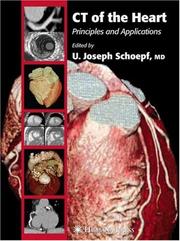
The introduction of fast ECG-synchronized computed tomography (CT) techniques enables imaging of the heart with a combination of speed and spatial resolution unparalleled by other noninvasive imaging modalities. Applying these modalities for the evaluation of coronary artery disease is a topic of active current research. Coronary artery calcium measurements are investigated as a marker for cardiac risk stratification. With contrast-enhanced CT coronary angiography, coronary arteries can be visualized with unprecedented detail, so that noninvasive stenosis assessment appears within reach. With increasing accuracy CT enables evaluation of coronary artery bypass grafts and stents. The cross-sectional nature of CT may to some degree allow noninvasive assessment of the coronary artery wall. CT for evaluating cardiac perfusion, motion, and viability is being investigated. In CT of the Heart, leading radiologists, cardiologists, physicists, engineers, and basic and clinical scientists from around the world survey the full scope of current developments, research, and scientific controversy regarding principles and applications of cardiac CT. Richly illustrated with numerous black-and-white and color images, the book discusses the interpretation of CT of the heart in a variety of clinical, physiologic, and pathologic applications. The authors emphasize current state-of-the-art uses of computed tomography, but also examine emerging developments at the horizon. They review the technical basis of CT image acquisition as well as the tools for image visualization and analysis. Meticulous and comprehensive, CT of the Heart authoritatively defines the current status of computed tomography of the heart, offering a truly balanced view of its technology, applications, significance, and future potential.
Heart --- Tomography, X-Ray Computed. --- Coeur --- radiography. --- Tomography. --- Tomographie --- Heart -- Tomography. --- Diagnostic Imaging --- Radiographic Image Enhancement --- Cardiovascular System --- Tomography, X-Ray --- Image Interpretation, Computer-Assisted --- Diagnostic Techniques and Procedures --- Image Enhancement --- Anatomy --- Tomography --- Photography --- Diagnosis --- Analytical, Diagnostic and Therapeutic Techniques and Equipment --- Tomography, X-Ray Computed --- Radiography --- Medicine --- Health & Biological Sciences --- Cardiovascular Diseases --- Radiology, MRI, Ultrasonography & Medical Physics --- Cardiology. --- Cardiographic tomography --- Diseases --- Medicine. --- Radiology. --- Medicine & Public Health. --- Imaging / Radiology. --- Radiological physics --- Physics --- Radiation --- Clinical sciences --- Medical profession --- Human biology --- Life sciences --- Medical sciences --- Pathology --- Physicians --- Internal medicine --- Radiology, Medical. --- Clinical radiology --- Radiology, Medical --- Radiology (Medicine) --- Medical physics
Book
ISBN: 1848826494 9786612928222 1848826508 1282928228 Year: 2010 Publisher: London : Springer London : Imprint: Springer,
Abstract | Keywords | Export | Availability | Bookmark
Loading...Choose an application
- Reference Manager
- EndNote
- RefWorks (Direct export to RefWorks)
The comprehensive assessment of cardiovascular structure and function with computed tomography (CT) has progressed at an astounding rate due to advances in scanning technology and image processing. Given the growing importance of cardiovascular CT, this book collates all relevant imaging findings and presents them in a clinically relevant and practical manner appropriate for the spectrum of physicians who diagnose and treat cardiovascular disease. The chapters have been written by an internationally renowned group of contributing authors and present discussion and images which characterize the full spectrum of cardiovascular CT.
Coronary Angiography -- Methods. --- Coronary Artery Disease -- Radiography. --- Heart -- Tomography. --- Tomography, X-Ray Computed -- Methods. --- Heart --- Cardiovascular system --- Diseases --- Diagnostic Imaging --- Tomography, X-Ray --- Radiographic Image Enhancement --- Image Interpretation, Computer-Assisted --- Investigative Techniques --- Methods --- Cardiovascular Diseases --- Tomography, X-Ray Computed --- Radiography --- Cardiac Imaging Techniques --- Analytical, Diagnostic and Therapeutic Techniques and Equipment --- Image Enhancement --- Diagnostic Techniques and Procedures --- Tomography --- Photography --- Diagnosis --- Medicine --- Health & Biological Sciences --- Tomography. --- Diagnosis. --- Cardiographic tomography --- Medicine. --- Radiology. --- Internal medicine. --- Cardiology. --- Cardiac surgery. --- Medicine & Public Health. --- Imaging / Radiology. --- Diagnostic Radiology. --- Internal Medicine. --- Cardiac Surgery. --- Radiology, Medical. --- Surgery. --- Cardiac surgery --- Open-heart surgery --- Medicine, Internal --- Clinical radiology --- Radiology, Medical --- Radiology (Medicine) --- Medical physics --- Internal medicine --- Surgery --- Radiological physics --- Physics --- Radiation
ISBN: 1280727098 9786610727094 3540495460 3540255230 Year: 2007 Publisher: Berlin, Heidelberg : Springer Berlin Heidelberg : Imprint: Springer,
Abstract | Keywords | Export | Availability | Bookmark
Loading...Choose an application
- Reference Manager
- EndNote
- RefWorks (Direct export to RefWorks)
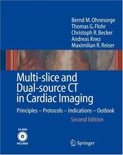
Preface to the Second Edition Despite worldwide efforts to assess and control cardiovascular risk factors, cardiac diseases and in particular coronary artery disease (CAD) are still the foremost causes of death in the developed countries of Western Europe, North America and Asia and are becoming increasingly common in Eastern Europe and the developing world (Deanfield 2001). Approximately one in five deaths is currently related to cardiac disease in Europe and the US. Nearly 500,000 deaths caused by CAD are reported every year in the US, over 600,000 in Europe, 170,000 of these in Germany alone. Over 12 million US citizens have a history of CAD, while every year 1. 1 million US and 300,000 German citizens suffer a coronary attack and more than 40% will die as a result of these attacks. Every second patient dies without prior symptoms and, in most cases, myocardial infarction occurs without warning. Once a blockage of the coronary arteries has occurred, death may ensue within a few minutes, even before hospitalization is possible. These alarming statistics highlight an acute need for tools to diagnose cardiac and coronary artery disease. Presently, the gold-standard mod- ity for diagnosis of CAD is invasive selective coronary angiography. More than 2.
Heart --- Imaging. --- Diseases --- Diagnosis. --- Cardiac diagnostic imaging --- Cardiac imaging --- Diagnostic cardiac imaging --- Imaging of the heart --- Imaging --- WG 141.5 Cardiovascular Diseases, Diagnosis and Therapeutics -- Specific diagnostic methods --- Diagnosis --- Heart Diseases --- Tomography, X-Ray Computed --- Radiology, Medical. --- Cardiology. --- Imaging / Radiology. --- Internal medicine --- Clinical radiology --- Radiology, Medical --- Radiology (Medicine) --- Medical physics --- Radiology. --- Radiological physics --- Physics --- Radiation
Book
ISBN: 3319425757 3319425730 Year: 2019 Publisher: Cham : Springer International Publishing : Imprint: Springer,
Abstract | Keywords | Export | Availability | Bookmark
Loading...Choose an application
- Reference Manager
- EndNote
- RefWorks (Direct export to RefWorks)
This volume provides a comprehensive and up-to-date account of the use of MRI and CT to identify and characterize developmental anomalies and acquired diseases of the female genital tract. Both benign and malignant diseases are considered in depth, and detailed attention is also paid to normal anatomic findings and variants. Further individual chapters focus on the patient with pelvic pain and the use of MRI for pelvimetry during pregnancy and the evaluation of fertility. Compared with the first edition, chapters have been either newly written by different authors or updated to reflect intervening progress; in addition, imaging of the placenta is now covered. Throughout, emphasis is placed on the most recent diagnostic and technical advances, and the text is complemented by many detailed and informative illustrations. All of the authors are acknowledged experts in diagnostic imaging of the female pelvis, and the volume will prove an invaluable aid to everyone with an interest in this field.
Pelvis --- Magnetic resonance imaging. --- Diseases --- Diagnosis. --- Radiology, Medical. --- Gynecology. --- Oncology . --- Diagnostic Radiology. --- Oncology. --- Tumors --- Gynaecology --- Medicine --- Generative organs, Female --- Clinical radiology --- Radiology, Medical --- Radiology (Medicine) --- Medical physics --- Genital Diseases, Female --- Magnetic Resonance Imaging --- Tomography, X-Ray Computed --- diagnostic imaging. --- methods. --- Radiology. --- Gynecology . --- Radiological physics --- Physics --- Radiation
Book
ISBN: 9788847008328 884700831X 9788847008311 9786612458866 1282458868 8847008328 Year: 2008 Publisher: Milano : Springer Milan : Imprint: Springer,
Abstract | Keywords | Export | Availability | Bookmark
Loading...Choose an application
- Reference Manager
- EndNote
- RefWorks (Direct export to RefWorks)
MDCT: From Protocols to Practice tackles contemporary and topical issues in MDCT technology and applications. As an updated edition of MDCT: A Practical Approach, this volume offers new content as well as revised chapters from the previous volume. New chapters discuss important topics such as imaging of children and obese subjects, the use of contrast medium in pregnant women coronary, MDCT angiography, and PET/CT in abdominal and pelvic malignancies. Furthermore an Appendix with over 50 updated MDCT scanning protocols completes this publication. The book emphasizes the practical aspects of MDCT, making it an invaluable source of information for radiologists, residents, medical physicists, and radiology technologists in everyday clinical practice.
Medicine & Public Health. --- Imaging / Radiology. --- Diagnostic Radiology. --- Interventional Radiology. --- Medicine. --- Medical radiology --- Interventional radiology. --- Médecine --- Radiologie médicale --- Radiologie interventionnelle --- Kalra, M. K. (Mannudeep K.). --- Tomography, Emission. --- Tomography, X-Ray Computed. --- Tomography, X-Ray Computed --- Tomography, X-Ray --- Image Interpretation, Computer-Assisted --- Radiographic Image Enhancement --- Image Enhancement --- Radiography --- Diagnostic Imaging --- Tomography --- Diagnostic Techniques and Procedures --- Photography --- Diagnosis --- Analytical, Diagnostic and Therapeutic Techniques and Equipment --- Radiology, MRI, Ultrasonography & Medical Physics --- Medicine --- Health & Biological Sciences --- 608.5 --- Computer tomografie --- CT --- Radiologie --- Diagnostic imaging. --- Clinical imaging --- Imaging, Diagnostic --- Medical diagnostic imaging --- Medical imaging --- Noninvasive medical imaging --- Computerized emission tomography --- Emission tomography --- PET (Tomography) --- PET-CT (Tomography) --- Positron emission tomography --- Positron emission transaxial tomography --- Radionuclide tomography --- Scintigraphy, Tomographic --- Tomography, Radionuclide --- Radiology. --- Diagnosis, Noninvasive --- Imaging systems in medicine --- Diagnostic imaging --- Positrons --- Radioisotope scanning --- Data processing --- Emission --- Radiology, Medical. --- Radiology, Interventional --- Therapeutics --- Clinical radiology --- Radiology, Medical --- Radiology (Medicine) --- Medical physics --- Interventional radiology . --- Radiological physics --- Physics --- Radiation
Book
ISBN: 3642017398 9786613087591 3642017401 1283087596 Year: 2011 Publisher: Berlin, Heidelberg : Springer Berlin Heidelberg : Imprint: Springer,
Abstract | Keywords | Export | Availability | Bookmark
Loading...Choose an application
- Reference Manager
- EndNote
- RefWorks (Direct export to RefWorks)
Dual-energy CT is a novel, rapidly emerging imaging technique which offers important new functional and specific information. With implementation of the technology in commercially available scanners, many clinical applications are now feasible. In this book, physicists and specialists from different CT manufacturers provide an insight into the technological basis of, and the different approaches to, dual-energy CT. Renowned medical scientists in the field explain the pathophysiological and molecular background of the technique, discuss its applications, provide detailed advice on how to obtain optimal results, and offer hints regarding clinical interpretation. The main focus is on the use of dual-energy CT in daily clinical practice, and individual sections are devoted to imaging of the vascular system, the thorax, the abdomen, and the extremities. Evaluations and recommendations are based on personal experience and peer-reviewed literature. Plenty of carefully chosen high-quality images are included to illustrate the clinical benefits of the technique.
Angiography. --- Cardiology. --- Dual-Source-Computertomographie. --- Internal medicine. --- Radiology, Medical. --- Tomography, X-Ray Computed -- Trends. --- Tomography. --- Dual energy CT (Tomography) --- Tomography, X-Ray --- Radiographic Image Enhancement --- Image Interpretation, Computer-Assisted --- Diagnostic Imaging --- Radiography --- Image Enhancement --- Tomography --- Photography --- Diagnostic Techniques and Procedures --- Diagnosis --- Analytical, Diagnostic and Therapeutic Techniques and Equipment --- Tomography, X-Ray Computed --- Medicine --- Health & Biological Sciences --- Radiology, MRI, Ultrasonography & Medical Physics --- Body section radiography --- Computed tomography --- Computerized tomography --- CT (Computer tomography) --- Laminagraphy --- Laminography --- Radiological stratigraphy --- Stratigraphy, Radiological --- Tomographic imaging --- Zonography --- Medicine. --- Radiology. --- Angiology. --- Medicine & Public Health. --- Imaging / Radiology. --- Diagnostic Radiology. --- Internal Medicine. --- Cross-sectional imaging --- Radiography, Medical --- Geometric tomography --- Blood-vessels --- Diagnosis, Radioscopic --- Heart --- Internal medicine --- Medicine, Internal --- Clinical radiology --- Radiology, Medical --- Radiology (Medicine) --- Medical physics --- Diseases --- Radiological physics --- Physics --- Radiation --- Diseases. --- Angiology --- Vascular diseases
| Listing 1 - 10 of 57 | << page >> |
Sort by
|